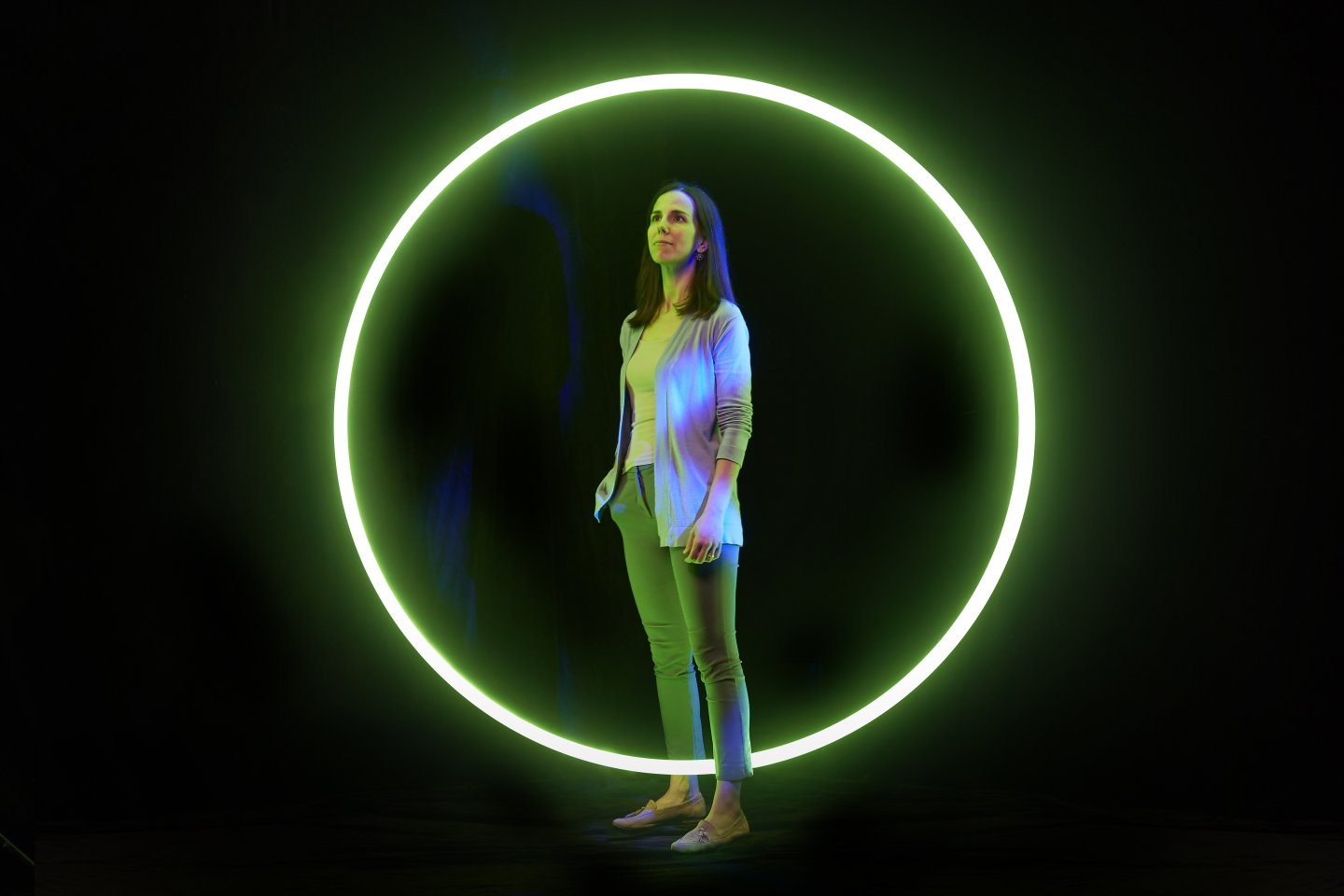
From particle collisions to data constellations
Dr Mireia Crispin-Ortuzar swapped her research on supersymmetry for a chance to reduce mortality from ovarian cancers.
Where were you when the Higgs Boson was discovered? For Dr Mireia Crispin-Ortuzar, the answer is easy. Right there, on the spot. A particle physicist by training, she was working at the Large Hadron Collider (LHC) in CERN when, in 2012, the ultimate ‘needle in a particle physics haystack’ issue was solved.
“It was such an exciting time; we thought we were going to find answers to all the major questions,” she recalls. “I was part of the ATLAS Collaboration, searching for particles that could explain supersymmetry and dark matter. Spoiler: we didn’t find anything. Well, we made many interesting measurements, but found no evidence of new particles.”

In common with Star Trekkers and The X-Files’ Mulder and Scully, Crispin-Ortuzar began to wonder if there was something else out there. Not other galaxies, or black holes, but something more concrete, here on Earth.
“I was doing my PhD, and at the same time organising conferences for undergraduate women in physics and other policy events,” she explains. “I began to think about the implications of science and technology for society, and I found myself starting to think about science in a different way.
“I loved the collaborative aspect of the LHC – thousands of scientists working together – but I began to feel I wanted to do something important for communities I was part of. I do think basic science is essential, but I wanted to do something more directly impactful for people.”
What if you could analyse images from people diagnosed with cancer, using smart, systemic computations to build better models than we have now?
At the same time, machine learning was beginning to become popular, she says. “I began reading academic papers on it, and thinking about how I might apply my skills in that area. It struck me that analysing data sets – millions of particle collisions – to find connections in order to answer different questions was exactly what I had been doing at CERN. And that’s what AI and machine learning does.”
From the stack of academic papers she read, medical imaging stood out. “Medicine was an obvious area with a lot of unanswered questions: around disease, and particularly cancer. I felt close to medical imaging, because PET and CT scans are basically mini versions of particle detectors. I thought this was a field I could really explore.
“Around this time there were a few papers published about applying machine learning to large data sets of medical images, and one mentioned the possibility of correlation with underlying biology, using molecular data from these same patients. I thought, ‘Oh my goodness, what if you could analyse images from people diagnosed with cancer, using smart, systemic computations to build better models than we have now, and also bring in the underlying biology? That could be really powerful.’”
In 2014, she applied for a Junior Research Fellowship (JRF) at Cambridge – a programme renowned for offering academic freedom and resources. “My application was 90% about particle physics, but I added a paragraph about applying it to medical imaging.” With the idea niggling away at her, she also wrote an unsolicited email to the then Head of Medical Physics at the Memorial Sloan Kettering Cancer Center in New York. “I wrote that I thought I could apply particle physics principles to medical imaging and cancer modelling. He invited me on a Skype call, and then eventually offered me a job as a postdoctoral researcher in his team. At almost the same time, I was offered the JRF at Trinity College. I asked Trinity: ‘Please could I go to New York first?’ Thankfully, they said yes.”
By the time she returned from the States, Crispin-Ortuzar was sold on radiology and, given the flexibility of the Fellowship, was able to choose to work on data integration for cancer, focusing on medical imaging. “I chose radiology for two reasons,” she says. “One: the potential impact is huge – everyone gets a scan at some point, so potentially everyone can benefit from the research. Two: it’s under-explored. Researchers tend to focus on genomics, combined with clinical data and perhaps pathology imaging. Radiology has been widespread since the early 20th century and these days it is sometimes perceived as less fashionable. But the beauty of it is that it’s a typical part of diagnostics and follow up, so there is so much information available. The range of scans makes studying changes in patients over time possible.
Thinking from the start about how to translate what you’re doing into clinical practice is inspiring.
“The measurements, however, have always been quite basic; they’re done by a radiologist in clinic who doesn’t have much time and is basically using a digital ruler to measure a lesion. It’s semi-quantitative at best. But adding computation offers so much potential: to see in 3D volumes; or to look at textures within the lesions and variations in the intensity of their colour (well, they’re black and white but the intensity of the grey). You can apply deep learning to quantify what you see and use it as part of a model.”
This is what she is doing now in her Computational Cancer Medicine lab: using radiology to explore the biology behind the patterns in the data, to understand why one cancer patient responds to treatment when another doesn’t.
“For example, we recently published a paper exploring neoadjuvant therapy – a kind of chemotherapy – in ovarian cancer patients. Ovarian cancer is nearly always metastatic – spreading throughout the body by the time it’s diagnosed, because its symptoms are so vague. The therapy only works on 60% of women, it can have nasty side effects and, if it doesn’t work, you may have wasted crucial time. “Our study used radiology to extract the textural features of the tumour, including from the healthy area immediately around it, and combined this data with clinical information and demographics including previous treatments, biomarkers and circulating tumour DNA from elsewhere in the body. The model we built showed that adding the radiological data gave us the greatest insight into what was going on. Now we’ll refine the model and the imagery, using techniques such as cellular microscopy and spatial sequencing, and we’ll improve the answers.”
Crispin-Ortuzar’s desire for direct impact is also fulfilled in her startup, 52 North, founded with three other Cambridge graduates, where she is Chief Digital Officer. The interdisciplinary team has created a low-cost device that can identify chemotherapy patients at risk of developing neutropenic sepsis, a side effect of the treatment that can ultimately lead to death. Designed with patients, it is now going into clinical trials. “Academia is focused on conferences, papers, study results,” she reflects. “A startup focuses on deliverables, which is beneficial for an academic. Thinking from the start about how to translate what you’re doing into clinical practice is inspiring.”
And it’s no accident that Crispin-Ortuzar is studying ovarian rather than another kind of cancer. She co-leads the Ovarian Cancer Programme at the CRUK Cambridge Centre and hopes to expand it to include other gynaecological cancers. “Ovarian cancer, like other gynaecological cancers, such as endometrial – is under-studied. We need data-driven approaches to solving these challenges, better collaborations, large-scale trials and robust discoveries. Alongside other women in my field, I feel I have a duty to do something about it.
“If you look at the data on funding versus mortality rates for different cancers, ovarian cancer is near the bottom. We know from the massively improved survival rate for breast cancer that if we put the work and resources into something, we can solve it.”
Dr Mireia Crispin-Ortuzar is Assistant Professor at the Department of Oncology and group leader at the Early Cancer Institute. To find out more about Cambridge cancer research, please contact Mary-Jane Boland.
 CAM
CAM

